
BioGPU The cellular biology runtime for AI agents
AI agents become experimental cell biologists. On BioGPU they design and run experiments inside cellular networks, see the molecular activities that respond in disease-relevant cells, and use that evidence to design better therapeutics.
Already built. Used in 25 drug-discovery programs.
Anima has built BioGPU, the cellular biology runtime for AI agents: a foundation model-driven experimental platform developed over more than a decade.
Built through pharmaceutical collaborations
BioGPU was developed with over $150 million received through strategic pharmaceutical collaborations with Lilly, Takeda and AbbVie.



What AI is missing
AI agents are blind to biology. With BioGPU, they see it.
Existing knowledge helps AI formulate biological hypotheses. Determining what a new intervention actually does inside a diseased cell requires new measurements. On BioGPU, AI agents design and run cellular network experiments that return evidence for testing hypotheses and guiding therapeutic design.
The evidence
A leading AI got the biology wrong. BioGPU showed what actually happened.
We asked it where a protein goes after a cancer drug. From the literature it answered with 80% confidence, and was wrong. BioGPU's data showed what the cells actually did.
The questionWhere does TFEB, the switch for the cell's recycling system, go 24 hours after the PI3K/mTOR cancer drug Gedatolisib, in four human cell states?
 Untreated · spread out
 After the drug · specks
What it means: an assay built to watch the nucleus would have called a working drug inactive.
- 3–4 of 30+committed predictions held
- 9–12could have misdirected a program
Across two internal runs.
How AI learns biology
Morphology shows that a cell changed. BioGPU shows which molecular activity changed.
That is the evidence AI needs to learn biology and design drugs against it.
- Cell Painting · morphologySees that the cell's appearance changed.Not which molecular activity changed.
- Single assaysMeasure selected molecules, assay by assay.Not the network around it.
- Lab-in-the-loopRuns the experiment the AI asks for.Returns its standard readouts.
- BioGPUSees which selected molecular activities changed, measured together in the same cells.Returns them to the AI as Cellular Biology Tokens it can reason on.
Complementary methods, described by typical use. How we test what the tokens add →
Applications
Every drug acts on a network. Now AI can design against it.
Binding and molecular properties do not by themselves describe a compound's cellular network effect. BioGPU brings measurements of selected molecular activities into the design loop, for every molecule AI designs. The resolved network also informs target and mechanism decisions before the first molecule is made.
Example · antibodies
After binding, a drug's biology begins. BioGPU brings it into the design loop.
Industry standard todayAI designs the antibody and validates binding.
With BioGPUWhat the binding does across the cellular network.
The same loop applies across modalities. All therapeutic workflows →
See the pharma workflow → See available targets → Explore cellular systems →
How it works
Reason. Run. Recognize. Resolve.
BioGPU is an operating experimental system, not an image collection: molecular visualizers, disease-relevant cellular systems, robotics, the Visual Biology Foundation Model, AI agents and Anima's scientists, working together. Built over more than a decade, it connects the design of a biological experiment to the interpretation of its results, returned to the agent as Cellular Biology Tokens.
- 01ReasonAI agents reason on the network state and design the next cellular network experiment.
- 02RunAI agents design and run the experiment: the BioGPU Agent selects the molecular-specific visualizers and perturbations in the program's cellular system. BioGPU's robotic lab executes it in parallel across conditions, perturbations and timepoints, in disease-relevant cells, including patient-derived cells where the program uses them.
- 03RecognizeThe Visual Biology Foundation Model recognizes the selected molecular activities, such as protein interactions, translocation, translation, RNA localization and signaling states, not just changes in overall cell appearance.
- 04ResolveCellular Biology Tokens return the result, and the agents design the next experiment. The loop repeats, and each round resolves more of the network around the target.
Cellular Biology Tokens
Cell biology, in a form AI can reason on.
BioGPU returns Cellular Biology Tokens that connect experimental images, biological interpretation and experimental context, including metadata and embeddings. Agents examine what changed, under which conditions, and with what supporting observations, then design the next experiment.
The core technology
A foundation model that knows what cell biology looks like.
Its training data did not exist before Anima produced it. Anima owns that image corpus and the Visual Biology Foundation Model trained on it.
Anima's Visual Biology Foundation Model has learned the visual signatures of protein interactions, translocation, translation, RNA localization and many more. Every run of a program adds Cellular Biology Tokens and tests the agent's predictions against what was measured.
Patient-derived cells
Real disease biology, including in cells from patients.
Each BioGPU program runs in cellular systems chosen for its disease biology. Where a program calls for it, that system uses cells derived from patients, so the network AI works on carries the disease itself. Primary, iPSC-derived and engineered systems widen the range, and new ones can be purchased or brought by you.
BioGPU in action
Resolving the network a drug acts on, so AI can design better drugs.
Three disease programs run on BioGPU.
 Normal bronchial
 Lung cancer
Resolving the cellular network around MYC, to optimize compounds against it.
Discuss this program → Healthy
 ALS
Resolving the cellular network around SOD1, to find a new way to modulate an undruggable target.
Discuss this program → Healthy donor
 RA patient
Resolving the disease network in patient-derived cells.
See the case →Team
Integrated capabilities across every discipline BioGPU needs.
Cell biology, imaging, chemistry, omics, systems biology, AI, software and drug discovery, integrated in one company.
Leadership
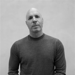
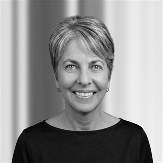
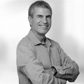
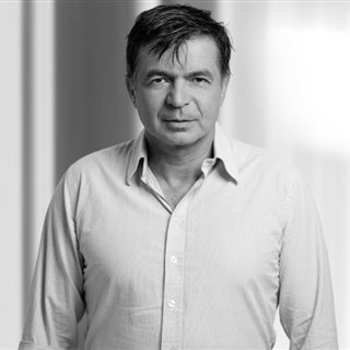
Yochi Slonim M.Sc., Co-founder & CEO, Board member
Iris Alroy PhD., Co-founder & CSO
Avi Eliassaf Chief Operating Officer
Yossi Oulu M.Sc., Vice President, Software Development
Generoso Ianniciello M.Sc., Chief Business Officer
Give your AI the biology it is missing.
Connect drug design with functional biology, for better drugs. Your own agents plug straight into BioGPU through its Runtime Skills and run their own workflows there.
Pharma team? See how your AI connects →
Reason · Run · Recognize · Resolve
Diagrams on this page are schematic. The cell images in The evidence and in the two Cellular Biology Tokens are measured data.